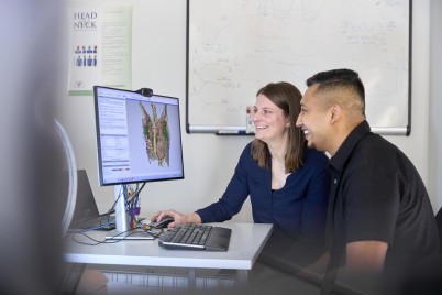
Dr Hayley Reynolds and Dr Tharanga Don
After receiving a Health Research Council fellowship in 2020, Dr Hayley Reynolds established a Cancer Imaging research group at the Auckland Bioengineering Institute (ABI), University of Auckland, following seven years working in Australia. She then set about developing new tools to help clinicians make better decisions for their cancer patients and improve treatment outcomes.
Her fellowship work has now led to the development of interactive software using large medical imaging databases to predict how melanoma and breast cancer could spread throughout the lymphatic system.
“With the bioengineering tools that we have at the ABI – including capabilities to develop computational models of the human body – we were able to map what was found in thousands of medical imaging records onto a computational model to present it in a more informative way for clinicians,” says Dr Reynolds.
She and her team mapped the melanoma imaging data from over 19,000 patients onto a 3D computer model of the skin and lymph nodes. The mapped information is presented as a web tool so that clinicians and patients can click on the 3D model and chose a region of skin, with the software able to show the predicted spread of cancer through the lymphatic system. This tool will be publicly available later this year following publication of the study results.
“These tools could help doctors plan surgery and radiation therapy more accurately, providing more personalised treatment and reducing potential side effects.”
They developed a similar tool for breast cancer to predict the potential spread of cancer to the lymphatic system using data from more than 860 patients and this is now freely available to clinicians. This work has attracted international interest, particularly for its potential application in radiation therapy planning and as a medical education tool.
“These tools could help doctors plan surgery and radiation therapy more accurately, providing more personalised treatment and reducing potential side effects,” says Dr Reynolds.
These new melanoma and breast cancer software tools are also being used in another Marsden-funded study where Dr Reynolds is creating computational models of the lymphatic system to better understand how and why lymphedema – a common side effect of cancer treatment – occurs.
“Lymphedema is a chronic swelling that often arises, especially in the legs and arms, when the lymphatic system is compromised. At this stage, there is no way to confidently predict if someone will get it and no definitive way to cure it,” says Dr Reynolds.
“We’re collaborating with researchers at the Australian Lymphoedema Education, Research and Treatment (ALERT) Centre at Macquarie University in Sydney, Australia, to develop new imaging devices and predictive tools to better visualise the lymphatic system and understand patterns of dysfunction in lymphedema.”
As part of the HRC fellowship, Dr Reynolds has also completed a small pilot clinical trial with prostate cancer patients at Auckland Hospital who were undergoing a particular type of radiation treatment called SABR (stereotactic ablative radiotherapy), where they receive higher doses of radiation in fewer treatments than conventional radiotherapy. Her team used advanced MRI scans taken prior to treatment and then six months and 12 months post-treatment to identify imaging biomarkers that could detect when radiation therapy may not be working.
“We’ve been trying to understand what changes we see in those MRI scans so we can tailor radiation therapy more precisely and enable doctors to offer salvage treatments sooner if a patient is not responding, giving patients the best chance of successful treatment. This is part of a larger project to deliver biologically guided radiation therapy, which aims to personalise radiation therapy rather than treating everyone in the same way.”
“Our goal was to work out if there are biomarkers within those images that we can assess at the six-and-12-month mark that will tell us if the patient will be cured or will need further treatment.”
Based on promising findings from the pilot trial, Dr Reynolds is working with radiation oncologists to expand the trial across New Zealand and Australia, with the aim of validating and then translating these biomarkers into clinical use. Findings include identifying some promising biomarkers from images that measure tumour cellularity and hypoxia (a known cause of resistance to radiation therapy).
“These predictive software and imaging biomarkers will provide advanced tools to improve cancer management when patients are in the early stages of their disease, hopefully reducing the need for more expensive treatments when cancer is more advanced and more likely to be incurable,” says Dr Reynolds.
Useful links:
The breast cancer predictive software can be downloaded from: GitHub - jsit433/BreastCancerAtlasExtension. You can also read the paper about this tool in Cancer Imaging.
Cancer Imaging Group – University of Auckland
The HRC funded Dr Hayley Reynold’s research through a 2020 HRC Sir Charles Hercus Health Research Fellowship to the value of $470k. During this fellowship, Dr Reynolds trained and supervised six postgraduate students and one postdoctoral student.